您的位置:首页 > 产品中心 > Anti-CEACAM1/CD66a Antibody, clone B3-17
Anti-CEACAM1/CD66a Antibody, clone B3-17

产品别名
Anti-CEACAM1/CD66a Antibody, clone B3-17
Carcinoembryonic antigen-related cell adhesion molecule 1, Biliary glycoprotein 1, BGP-1, CD66a
基本信息
| eCl@ss | 32160702 |
| NACRES | NA.41 |
| General description【一般描述】 | CEACAM1 (carcinoembryonic antigen-related cell adhesion molecule 1) is thought to be a member of the immunoglobulin superfamily and is known as an epithelial tumor suppressor and an angiogenic growth factor. It has also been linked to the actin-based cytoskeleton. CEACAM1 is also known as a cellular receptor for a number of human mucosa pathogenic bacteria. The loss of activity of CEACAM1 has been related to the development of colorectal cancer. |
| Immunogen【免疫原】 | Recombinant human CEACAM1/CD66a. |
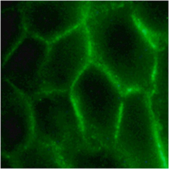
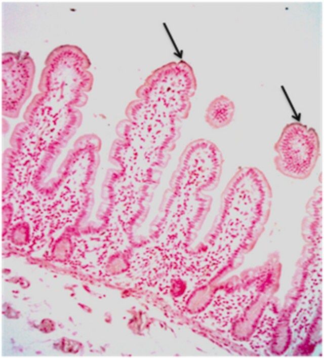
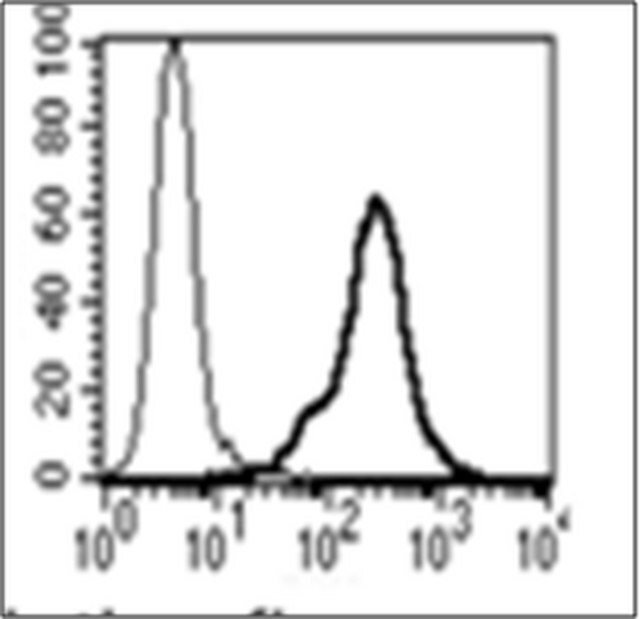
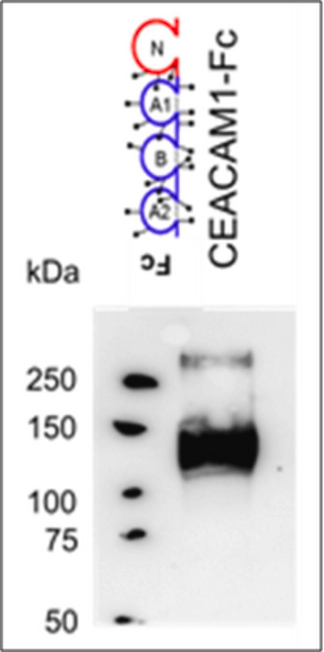
| Application【应用】 | ELISA Analysis: 10 µg/mL from a representative lot detected human CEACAM1/CD66a by ELISA (Courtesy of Dr. B. Singer, University Duisburg-Essen, Germany). Flow Cytometry Analysis: 10 µg/mL from a representative lot detected the exogenously expressed human CEACAM1/CD66a on the surface of transfected HeLa cells (Courtesy of Dr. B. Singer, University Duisburg-Essen, Germany). Immunocytochemistry Analysis: 10 µg/mL from a representative lot detected CEACAM1/CD66a on the surface of HT-29 human colorectal carcinoma cells (Courtesy of Dr. B. Singer, University Duisburg-Essen, Germany). Immunohistochemistry Analysis: 10 µg/mL from a representative lot detected CEACAM1/CD66a immunoreactivity in human jejunum tissue (Courtesy of Dr. B. Singer, University Duisburg-Essen, Germany). Western Blotting Analysis: 10 µg/mL from a representative lot detected the exogensously expressed human CEACAM1/CD66a extracellular domain-Fc fusion in lysates from transfected cells (Courtesy of Dr. B. Singer, University Duisburg-Essen, Germany). Flow Cytometry Analysis: Representative lots, either unconjugated or FITC-conjugated, detected CECAM1 immunoreactivity on the surface of human peripheral blood naïve and memory B-cells (Khairnar, V., et al. (2015). Nat. Commun. 6:6217; Seifert, M., et al. (2015). Proc. Natl. Acad. Sci. 112(6):E546-555). Flow Cytometry Analysis: A representative lot detected CEACAM1/CD66a induction on the surface of normal human bronchial epithelial (NHBE) cells stimulated with poly(I:C) or interferons (Klaile, E., et al. (2013) Respir Res. 14:85). Flow Cytometry Analysis: A representative lot detected CEACAM1/CD66a expression on the surface of 2 day-starved human epithelial (HT29, T102/3) and endothelial (AS-M.5) cells, as well as multivesicular bodies (MVBs) derived from these cells (Muturi H.T., et al. (2013) PLoS One. 8(9):e74654). Immunocytochemistry Analysis: A representative lot detected CEACAM1/CD66a immunoreactivity on the surface of normal human bronchial epithelial (NHBE) cells by indirect immunofluorescence staining (Klaile, E., et al. (2013) Respir Res. 14:85). Immunohistochemistry Analysis: A representative lot detected CEACAM1/CD66a in paraffin-embedded human lung cancer tissue sections (Klaile, E., et al. (2013) Respir Res. 14:85). Western Blot Analysis: A representative lot detected CEACAM1/CD66a in 2 day-starved AS-M.5 human endothelialn cells and AS-M.5-derived multivesicular bodies (MVBs) (Muturi H.T., et al. (2013) PLoS One. 8(9):e74654). This Anti-CEACAM1/CD66a Antibody, clone B3-17 is validated for use in ELISA, Flow Cytometry, Immunocytochemistry, Immunohistochemistry, and Western Blotting for the detection of human CEACAM1/CD66a. |
| Quality【质量】 | Evaluated by Western Blotting in HepG2 cell lysate. Western Blotting Analysis: 2 µg/mL of this antibody detected CEACAM1/CD66a in 10 µg of HepG2 cell lysate. |
| Physical form【外形】 | Protein G purified. Format: Purified |
| Other Notes【其他说明】 | Concentration: Please refer to lot specific datasheet. |
产品性质
| Quality Level【质量水平】 | 100 |
| biological source【生物来源】 | mouse |
| antibody form【抗体形式】 | purified immunoglobulin |
| antibody product type | primary antibodies |
| clone【克隆】 | B3-17, monoclonal |
| species reactivity | human |
| technique(s) | ELISA: suitable flow cytometry: suitable immunocytochemistry: suitable immunohistochemistry: suitable (paraffin) western blot: suitable |
| isotype【同位素/亚型】 | IgG1κ |
| NCBI accession no.【NCBI登记号】 | NP_001020083 |
| UniProt accession no.【UniProt登记号】 | P13688 |
| shipped in【运输】 | wet ice |
产品说明
| Target description【目标描述】 | ~160 kDa observed. Target band size appears larger than the calculated molecular weights of 57.56/53,80 kDa (pro-/mature isoform 1; BGPa, CEACAM1-4L, TM1-CEA), 45.95/42.19 kDa (pro-/mature isoform 2; BGPg, CEACAM1-4C1), 35.30/31.54 kDa (pro-/mature isoform 3; BGPh, CEACAM1-3), 38.58/34.81 kDa (pro-/mature isoform 4; BGPi, CEACAM1-3C2), 50.35/46.58 kDa (pro-/mature isoform 5; BGPy, CEACAM1-3AL), 46.91/43.15 kDa (pro-/mature isoform 6; BGPb, CEACAM1-3L, TM2-CEA), 27.43/23.67 kDa (pro-/mature isoform 7; BGPx, CEACAM1-1L), 50.52/46.76 kDa (pro-/mature isoform 8; BGPc, CEACAM1-4S, TM3-CEA), 43.06/39.30 kDa (pro-/mature isoform 9; BGPz, CEACAM1-3AS), 51.15/47.38 kDa (pro-/mature isoform 10), 39.87/36.11 kDa ((pro-/mature isoform 11; BGPd, CEACAM1-3S) due to glycosylation. Uncharacterized bands may be observed in some lysates. |
| 【免责声明】 | Unless otherwise stated in our catalog or other company documentation accompanying the product(s), our products are intended for research use only and are not to be used for any other purpose, which includes but is not limited to, unauthorized commercial uses, in vitro diagnostic uses, ex vivo or in vivo therapeutic uses or any type of consumption or application to humans or animals. |
安全信息
| Storage Class Code【储存分类代码】 | 12 - Non Combustible Liquids |
| WGK | WGK 1 |


