


您的位置:首页 > 产品中心 > PhosphoDetect Anti-Neurofilament H Mouse mAb (SMI-31)
PhosphoDetect Anti-Neurofilament H Mouse mAb (SMI-31)
产品性质
| Quality Level【质量水平】 | 100 |
| biological source【生物来源】 | mouse |
| antibody form【抗体形式】 | purified antibody |
| antibody product type | primary antibodies |
| clone【克隆】 | SMI-31, monoclonal |
| form【形式】 | liquid |
| contains【包含】 | ≤0.1% sodium azide as preservative |
| species reactivity | Xenopus, chicken |
| species reactivity (predicted by homology) | mammals |
| manufacturer/tradename | Calbiochem® |
| storage condition【储存条件】 | OK to freeze avoid repeated freeze/thaw cycles |
| isotype【同位素/亚型】 | IgG1 |
| shipped in【运输】 | wet ice |
| storage temp.【储存温度】 | −20℃ |
| packaging【包装】 | 100 μL in Glass bottle |
基本信息
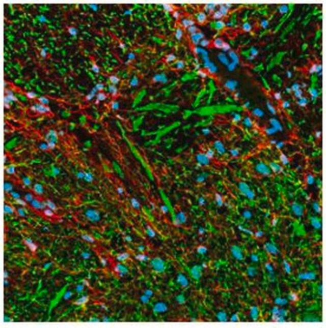
| General description【一般描述】 | This PhosphoDetect Mouse monoclonal antibody supplied as purified antibody. Recognizes the ~180-200 kDa phosphorylated neurofilament H protein. Recognizes the ~180-200 kDa phosphorylated neurofilament H protein in rat central nervous system (CNS) cytoskeletal preparations. Also weakly recognizes phosphorylated NF-M protein. |
| Immunogen【免疫原】 | homogenized, hypothalami from Fischer 344 rat brain |
| Application【应用】 |  ELISA (1:1000) Frozen Sections (1:1000, see comments) Immunoblotting (1:1000) Immunocytochemistry (1:1000, see comments) Paraffin Sections (1:1000, heat pre-treatment required, see comments) Immunoprecipitation (see comments) |
| Warning【警告】 | Toxicity: Standard Handling (A) |
| Physical form【外形】 | In PBS and Thimerosal |
| Other Notes【其他说明】 | Strongly recognizes phosphorylated neurofilament H and, to a lesser extent, phosphorylated neurofilament M. By immunocytochemistry this antibody broadly stains thick and thin axons and some dendrites, such as basket cell dendrites, but not Purkinje cell dendrites. Does not generally stain nerve cell bodies or other cells and tissues except peripheral axons. May also stain neuronal cell bodies in pathological conditions. Aberrant phosphorylation of neurofilament H in cell bodies can be demonstrated in neuronal cell cultures following treatment with agents that induce stress-activated protein kinase. This antibody is reported to co-immunoprecipitate neurofilament-associated kinase (NAK 115) via interaction of the antibody with the tail domain of neurofilament H. Phosphatase treatment of tissue sections or immunoblotting samples abolishes antibody reactivity. Reactivity is unaffected by trypsin treatment of samples. Tissues and cultured cells can be fixed with a variety of paraformaldehyde- or formaldehyde-containing fixatives, including Bouin′s fixative. Post-fixation in cold methanol or methanol/hydrogen peroxide facilitates access of the antibody to the epitope in frozen sections or thick tissue sections fixed in 4% paraformaldehyde and in cultured cells. For staining formalin-fixed, paraffin sections it is recommended that the de-paraffinized tissue be autoclaved in dH2O for 10 min or boiled in Tris-buffered saline, pH 9.0, in a microwave for 15 min to expose the epitope. Antibody should be titrated for optimal results in individual systems. Raina, A.K., et al. 1999. Neuroreport10, 1355. Yang, C.C., et al. 1998. Brain121, 1089. Giasson, B.I and Mushynski, W.E. 1996. J. Biol. Chem.271, 30404. Mirabella, M., et al. 1996. J. Neuropath. Exp. Neurol.55, 774. Xiao, J. and Monteiro, M.J. 1994. J. Neurosci.14, 1820. |
| Legal Information【法律信息】 | CALBIOCHEM is a registered trademark of Merck KGaA, Darmstadt, Germany |